Day 1 :
- Magnetic Resonance Imaging
Session Introduction
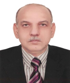
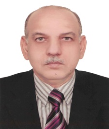
Musarrat Hasan
Institute of Ultrasound Imaging Karachi -Pakisatn
Title: Differential Diagnosis of Focal Masses in Liver with Special Reference to Tropical Disease

Biography:
Director & CEO Institute of Ultrasound Imaging since 1984, which is affiliated with JUREI at Thomas Jefferson University Hospital, Philadelphia PA, USA. Visiting Fellowship in Ultrasound from Thomas Jefferson University Hospital, Philadelphia USA 1982. Founder member & past President Ultrasound Society of Pakistan. Chairman History and Archiving AFSUMB. Fellow member, A I U M and recipient of 2001 Award for contributing & promoting Ultrasound. Has delivered more than 26000 hours of lectures / talks /scientific papers in ultrasound during last 33 years. Discovered New Contrast Agent for Tubal patency.
Abstract:
Liver focal masses may be seen in variety of clinical settings ranging from incidental findings to symptomatic patients or as apart of routine targeted scanning to look for hepatic tumor. Haemangioma, simple cyst Focal Nodular Hyperplasia are quite often seen. Hepatocellular Ca, and metastasis are also not rare to see. In south East Asia and specially in Indo-Pak we encounter many varieties of Amoebic and pyogenic Abscess along with a vast variety of Hydatid cyst and ascaris. The role of ultrasound in detecting such masses with state of art equipment has reach new heights and one can easily differentiate one mass from another. There are few masses which overlap in sonographic appearance and need further evaluation with Color Doppler to establish the diagnosis.
To determine the nature of mass we follow certain guidelines as to weather the mass is cystic, solid, complex, predominantly cystic, predominantly solid, hypo echoic, echogenic, heterogeneous, thick and thin wall, halo or without halo, single, multiple and whether it is smooth or irregular in outer contour. My talk will discuss in depth all the parameters with special reference to tropical diseases.
Musarrat Hasan
Institute of Ultrasound Imaging Karachi -Pakisatn
Title: Experience with Gaseous Spring Water as Contrast Agent in Tubal Patency Assessment

Biography:
Abstract:
- Ultrasound
Session Introduction
Mohammed Elmetwally
Institute of Reproductive Biology
Title: Noninvasive color Doppler blood flow indices of uterine and umbilical blood vessels during pregnancy: sheep is the experimental model
Biography:
Mohammed Elmetwally has completed his PhD at the age of 30 years from Hannover University of Veterinary Medicine in Germany and postdoctoral studies from Cornell University. He is assistant professor of Theriogenology, faculty of Veterinary Medicine, Mansoura University, Egypt. He has published more than 13 papers in reputed journals.
Abstract:
Sheep represents an appropriate animal model to study the uterine blood perfusion in women, because the similarities of uterine vascularization and existence of pelvic anastomoses of non pregnant sheep with that in women (1,2). The objective of the present study was to characterize blood flow in the maternal and fetal blood flow changes during pregnancy in sheep. Herein 18 pregnant sheep were used in the present study. The blood flow indices includes blood flow volume (BFV), Time averaged maximum velocity (TAMV), resistance index (RI), pulsatility index (PI), Time averaged mean velocity, impedance of blood flow (AB or S/D ratio), peak velocity of blood flow (PV) and blood flow acceleration (Accel). Non-invasive Color Doppler Ultrasound examinations were started 2 weeks (wk) after breeding, and continued at 2-wk intervals until parturition. Blood flow volume, TAMV, PS as well as PV were increased (P<0.0001) continuously until parturition. Also, TAMEAN increased with advancing pregnancy (P<0.0001) and decreased (P<0.0001) after wk. 18. The uterine artery RI, PI and S/D were progressively decreased until parturition. On the other hand, the blood flow acceleration velocity was increased until wk. 16 in sheep begin to decrease until birth. For umbilical blood flow significant increases (P<0.001) of BFV, TAMV and TAMEAN were recognized until W 19 of pregnancy in sheep foeti and then decreased (P<0.001) until parturition. Conversely, UM-PI, RI and PS/ED were decreased significantly ((P<0.002-0.01) until W 19 and then increased (P<0.01-0.0001 resp.). The umbilical artery BFV increased (P<0.0001) during the pregnancy from 7.27±0.82 ml/min at W 6 of gestation to 700.51±31.05 ml/min at W 19 and then significantly (P<0.0001) decreased to 350.561±72.15 ml/min at W 20. Absence of end diastolic velocity (EDV) of umbilical blood flow is recognized in in all examined fetuses between Week 4 and Week 12. In conclusion, the obtained results may guide the state of intrauterine fetal growth retardation in abnormal pregnancies.
- Advances in Medical Imaging
Session Introduction
Bahar Mahjoubi
CRRC, Iran university of Medical Sciences, Tehran
Title: Characteristics of endo anal ultrasonography in people with wanted and unwanted anal intercourse
Biography:
Bahar Mahjoubi has completed MD degree at the age of 25, and general surgery at the age of 30 in IUMS, fellowship of colorectal surgery in Sydney University and NSW University of Australia. now she is Professor of colorectal surgery At IUMS ,CRRC. she has published 53 papers.
Abstract:
Anal intercourse is rarely discussed in the scientific literature, because of concerns about the effect of many women anal intercourse on their anal sphincter. We decided to help clinicians are consulted in this regard by endo anal ultrasonography provide objective evidence.
We aimed to evaluate the presence of sphincter damage among women who referred from Legal institution and have complaints from anal intercourse (wanted or unwanted) with their partners.
we studied 40 female patients with mean age of 29.18+/_10.5 years old.All the patients underwent endo anal ultrasound by 360 degree/2D/3D B&K system by colorectal surgeon.
Eight of the patients (20%) had descent and 16 (40%) had internal prolapse.
Thirty two patients of 40 patients had gap in their external anal sphincter in endoanal ultrasound.
Among patients 17.5% had no external sphincter gap and 12.5% had gap site in 6→9 + 9→12 o’clock in lithotomy position.
Seven of patients (17.5%) had sphincter gap in both right and left side of the anal external sphincter.
Overall gap sites are more common in 12→6 o’clock of lithotomy position.the most common part of external sphincter disruption was in the 12→6 o’clock of lithotomy position.
According to data obtained, we are pleased that this abstract to be discussed in Conference of ultrasound to help guide the clinicians.
C Ganesh Pai
Department of Gastroenterology & Hepatology, Kasturba Medical College, Manipal University, Manipal 576104. Karnataka, India
Title: Endosonographic evidence of chronic pancreatitis according to Rosemont criteria in patients with unexplained recurrent acute pancreatitis

Biography:
Dr. C GANESH PAI is a professor and a full time teaching faculty at Kasturba Medical College, Manipal, India, heading the Department of Gastroenterology and Hepatology. His areas of research include chronic pancreatitis and inflammatory bowel diseases. He has received research grants from the Indian Council of Medical Research, Department of Biotechnology, Government of India and the Department of Science and Technology, Government of India.
Abstract:
Recurrent acute pancreatitis (RAP) is known to lead to CP. We aimed to assess the frequency of chronic pancreatitis in patients with unexplained RAP. Patients with RAP in whom no cause was evident on clinical and laboratory evaluation and on cross sectional imaging were prospectively evaluated by endocopic ultrasound (EUS) for changes of chronic pancreatitis (CP) using Rosemont criteria. Findings of endoscopic retrograde cholangiopancreatography (ERCP) as per Cambridge criteria were also considered in the subgroup in which this procedure was indicated for therapy. Forty-eight patients (male: female = 37:11, median age = 21, range 10 -70 years) with RAP seen over a 34-month period were included prospectively. All underwent EUS and 19 (39.6%) underwent ERCP additionally. EUS finding were most consistent for CP and suggestive of CP in 7 (14.6%) and 11 (22.9%) patients respectively. Two (4.2%) others developed findings most consistent with CP on repeat EUS during the study period. An additional 5 (10.4%) had mild or moderate changes of CP on ERCP. Thus, 25 (52.1%) patients showed findings most consistent with or suggestive of CP on EUS, or of mild to moderate changes of CP on ERCP during the study period. Our data show that more half of the patients without evidence of CP on other investigations show evidence of chronic disease on EUS or ERCP. This opens up the possibility of focusing on earlier stages of this condition to see if early interventions will alter the outcomes in these patients.
